Understanding the Condition
Not all tumor or abnormal tissue changes are cancerous. Many conditions treated with modern image-guided medicine are benign, meaning they do not spread to other parts of the body or behave like malignant cancers.
However, benign conditions can still significantly affect quality of life. Depending on their size, location and growth pattern, they may cause pain, pressure symptoms, reduced mobility, hormonal changes, or discomfort.
At Nordic Focal Therapies, we provide specialist assessment and minimally invasive image-guided treatment options for selected benign conditions. Using advanced CT and ultrasound imaging, our specialists can precisely evaluate abnormalities and determine the most appropriate management approach for each patient.
Our goal is to treat symptoms effectively while preserving healthy surrounding tissue whenever possible.
What Are Benign Conditions?
A benign condition refers to an abnormal growth or tissue change that is not cancerous.
Unlike malignant tumor, benign lesions generally:
Do not spread to distant organs
Grow more slowly
Remain localised to their original location
However, benign does not always mean harmless. Some benign conditions can cause problems because of their:
Size
Location
Pressure on nearby organs or nerves
Effect on movement or daily activities
Associated pain or discomfort
The decision to treat a benign condition depends on the individual situation, symptoms, and the impact on the patient’s quality of life.
Types of Benign Conditions We Evaluate and Treat
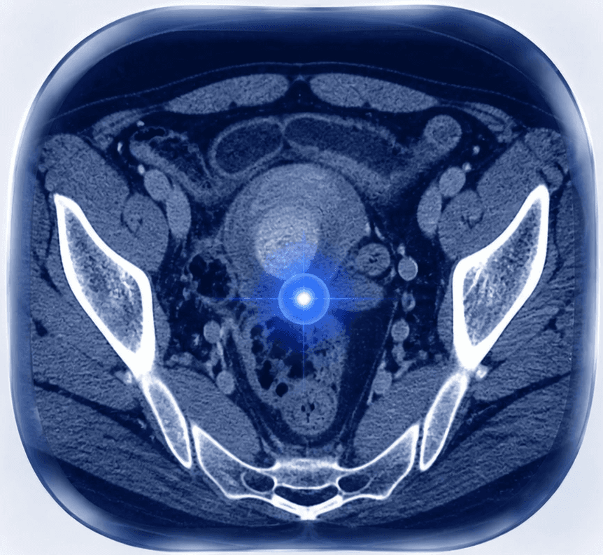
Uterine Fibroids (Myomas)
Uterine fibroids, also known as myomas, are non-cancerous growths that develop from the muscular tissue of the uterus.
They are very common and may not cause symptoms. However, larger or strategically located fibroids can affect daily life.
Possible symptoms include:
Heavy or prolonged menstrual bleeding
Pelvic pressure or discomfort
Lower abdominal pain
Increased urinary frequency due to pressure on the bladder
A feeling of fullness or abdominal enlargement
Treatment decisions depend on factors such as fibroid size, location, symptoms, age, and personal goals.
Selected symptomatic fibroids may be suitable for minimally invasive image-guided treatments.
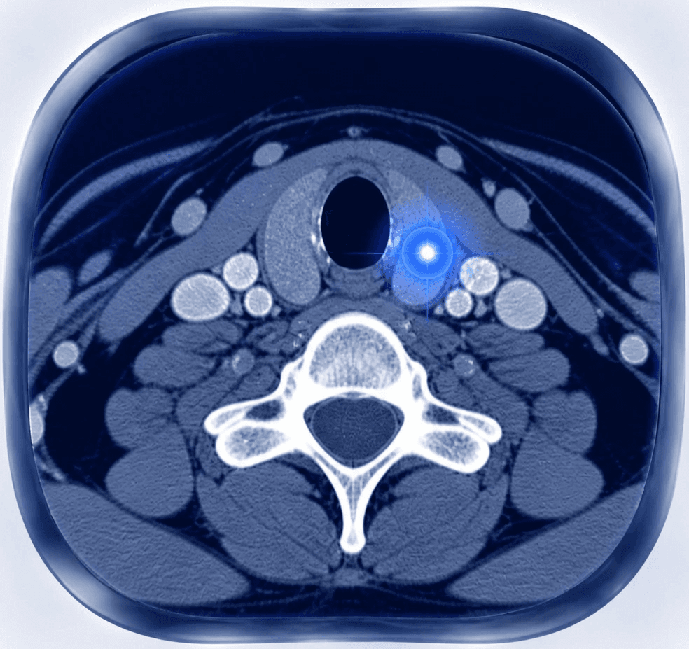
Thyroid Nodules and Benign Thyroid Enlargement
Thyroid nodules are common findings, and the majority are benign.
Many thyroid nodules require only monitoring, but some may grow and cause symptoms due to compression of nearby structures.
Possible symptoms include:
Visible enlargement in the neck
Feeling of pressure
Difficulty swallowing
Discomfort when wearing tight clothing
Cosmetic concerns
Evaluation typically includes ultrasound imaging and, when needed, tissue sampling.
For selected benign thyroid nodules, minimally invasive treatments may help reduce the size of the abnormal tissue while preserving thyroid function.
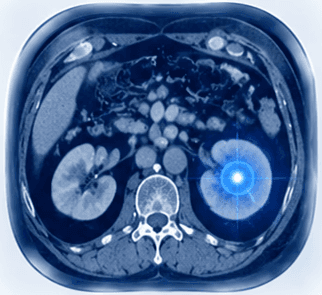
Desmoid Tumor
Desmoid tumor are rare benign soft tissue tumor that develop from connective tissue.
Although they do not metastasise like cancer, they can behave aggressively by growing into surrounding structures.
Depending on their location, desmoid tumor may cause:
Pain
Reduced movement
Pressure symptoms
Functional limitations
Because their behaviour varies between patients, management decisions require careful assessment of imaging findings, symptoms and tumour characteristics.
Endometriosis-Related Lesions
Endometriosis occurs when tissue similar to the lining of the uterus grows outside the uterus.
In some patients, endometriosis can form localised lesions or scar-related abnormalities that cause persistent pain.
Examples include scar endometriosis, which may develop after previous abdominal surgery.
Symptoms may include:
Localised pain
Tenderness around previous surgical scars
Pain that changes with the menstrual cycle
Discomfort affecting daily activities
Specialist assessment and imaging help determine the most appropriate treatment approach.
Osteoid Osteoma
Osteoid osteoma is a small benign bone tumour that most commonly affects children, adolescents and young adults.
Although it is not cancerous, it can cause significant pain.
Typical symptoms include:
Persistent localised bone pain
Pain that is often worse at night
Improvement with anti-inflammatory medication
Because these lesions are often very small, accurate CT imaging plays an important role in diagnosis and treatment planning.
Vascular Conditions
Some benign conditions involve abnormal veins or blood vessels.
Examples include:
Varicocele
Pelvic venous congestion
Venous malformations
These conditions may cause:
Chronic discomfort
Swelling
Pressure sensations
Pain affecting everyday activities
Image-guided vascular procedures can provide targeted treatment options for selected patients.
Symptoms of Benign Conditions
Symptoms vary widely depending on the type and location of the condition.
Possible symptoms include:
Localised pain
Pressure or fullness
Bleeding abnormalities
Reduced movement
Swelling or a palpable lump
Changes in appearance
Symptoms affecting nearby organs
Because benign and malignant conditions can sometimes appear similar on imaging, accurate diagnosis is essential.
How Benign Conditions Are Diagnosed
Diagnosis begins with a detailed assessment of symptoms, medical history and imaging findings.
Depending on the condition, evaluation may include:
Ultrasound
Ultrasound is often the first imaging method used for evaluating superficial structures, thyroid abnormalities, pelvic conditions and soft tissue lesions.
It provides real-time imaging without radiation and can also be used to guide minimally invasive procedures.
CT Imaging
CT provides detailed anatomical information and helps specialists evaluate:
Lesion size
Exact location
Relationship to surrounding structures
Treatment planning considerations
CT is particularly important for evaluating bone lesions and guiding certain procedures.
MRI
MRI provides excellent soft tissue detail and is frequently used for:
Soft tissue tumor
Endometriosis-related lesions
Complex anatomical areas
Musculoskeletal abnormalities
Image-Guided Biopsy
In some cases, imaging alone cannot confirm the exact nature of a lesion.
An image-guided biopsy allows specialists to obtain a small tissue sample using CT or ultrasound guidance.
The sample can help determine:
Whether a lesion is benign or malignant
The exact tissue type
The most appropriate management approach
How Benign Conditions Are Evaluated
Not every benign condition requires treatment.
Specialists consider:
Severity of symptoms
Impact on daily life
Size and location of the lesion
Growth over time
Previous treatments
Patient preferences
The goal is always to balance effective symptom relief with preservation of normal tissue and function.
The Role of Image-Guided Medicine
Advances in interventional radiology have created new options for selected patients with benign conditions.
Image-guided techniques allow specialists to:
Precisely locate abnormal tissue
Target treatment areas accurately
Minimise damage to surrounding healthy structures
Reduce the need for more invasive procedures in suitable cases
By combining advanced imaging with minimally invasive techniques, treatment can be tailored to each patient’s anatomy and needs.
When Should You Seek Specialist Assessment?
A specialist review may be appropriate if you:
Have a benign tumour causing symptoms
Have an uncertain lesion found during imaging
Experience persistent pain or pressure symptoms
Have been advised to consider surgery
Want to understand minimally invasive treatment options
A detailed review of your imaging and clinical situation can help identify the most appropriate next step.
Why Choose Nordic Focal Therapies?
Nordic Focal Therapies combines extensive university hospital experience with advanced image-guided minimally invasive medicine.
Our specialists have experience evaluating and treating both tumour-related and non-cancerous conditions using precise, image-guided approaches.
Our philosophy is:
Treat what matters. Preserve what is healthy.
Every patient is assessed individually, with treatment decisions based on imaging findings, symptoms, and personal goals.
Related Treatments
Depending on your condition, possible treatment options may include:
→ Image-Guided Tumour Treatments
→ Cryoablation
→ Microwave Ablation
→ Image-Guided Biopsies
→ Image-Guided Vascular Treatments
Frequently Asked Questions
Are benign tumor dangerous?
Most benign tumor do not spread or behave like cancer. However, they can still cause symptoms or affect surrounding structures depending on their size and location.
Do all benign conditions require treatment?
No. Many benign conditions can be safely monitored. Treatment is considered when symptoms, growth, or functional problems affect quality of life.
Can benign conditions be treated without surgery?
Some selected benign conditions can be treated using minimally invasive image-guided procedures instead of traditional surgery.
How do doctors know if a lesion is benign?
Diagnosis is based on a combination of imaging findings, clinical assessment and, when necessary, biopsy results.
What are the benefits of image-guided treatment?
Potential benefits include precise targeting, smaller treatment areas, reduced impact on surrounding tissue and shorter recovery compared with some surgical approaches.