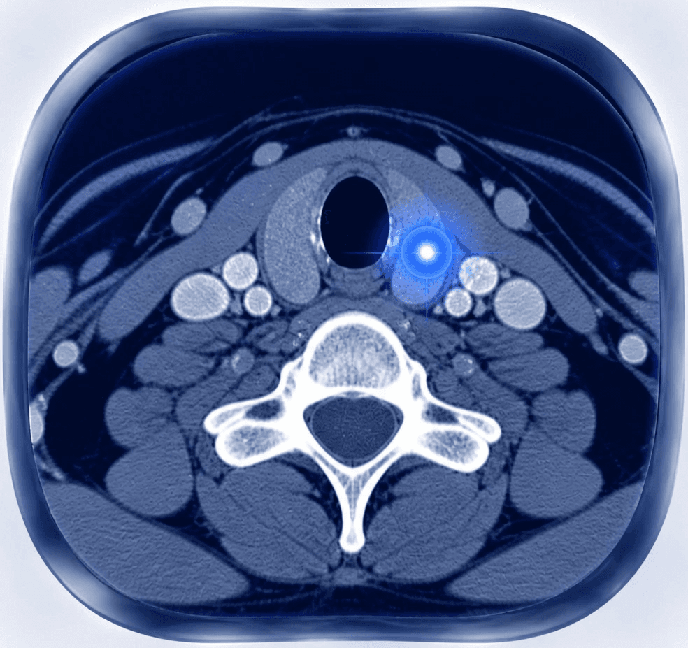
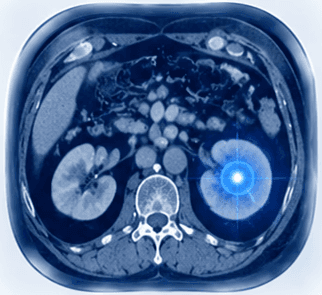
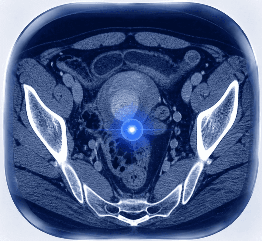
Understanding the Condition
Bones and soft tissues provide the structure, support and movement that allow the body to function. Like other tissues, they can be affected by different types of abnormalities, including benign lesions, primary tumor, and cancer that has spread from another part of the body.
At Nordic Focal Therapies, we specialise in image-guided assessment and minimally invasive treatments for selected bone lesions, soft tissue abnormalities and metastatic disease.
Using advanced CT and ultrasound imaging, our specialists can precisely evaluate abnormalities, obtain tissue samples when needed, and plan targeted treatments while protecting surrounding healthy structures.
What Are Bone and Soft Tissue Lesions?
A lesion is an area of abnormal tissue identified through imaging or clinical examination.
Lesions affecting bones and soft tissues can have many different causes, including:
Benign growths
Inflammatory changes
Primary bone or soft tissue tumor
Metastases from cancers elsewhere in the body
Determining the exact nature of a lesion is essential because different conditions require different approaches.
Modern imaging techniques allow specialists to evaluate these abnormalities with increasing accuracy and guide personalised treatment decisions.
Types of Conditions We Evaluate and Treat
Bone Metastases
Bone metastases occur when cancer cells spread from another part of the body to the bones.
They are common in advanced cancers, including:
Breast cancer
Prostate cancer
Lung cancer
Kidney cancer
Thyroid cancer
Other solid tumor
Bone metastases may affect different parts of the skeleton, including:
Spine
Pelvis
Ribs
Long bones
Depending on their location and symptoms, bone metastases can cause:
Persistent pain
Reduced mobility
Weakening of the bone structure
Increased risk of fracture
Modern cancer care often combines systemic treatments with local therapies aimed at controlling symptoms and improving quality of life.
Painful Bone Lesions
Not all painful bone lesions are caused by cancer.
Some benign bone conditions can also cause significant symptoms.
One example is:
Osteoid Osteoma
Osteoid osteoma is a small benign bone tumour that commonly affects younger patients.
Typical symptoms include:
Persistent localised pain
Night-time pain
Improvement with anti-inflammatory medication
Because these lesions can be very small, accurate imaging is essential for diagnosis and treatment planning.
Soft Tissue Tumor
Soft tissues include muscles, tendons, connective tissue, fat, and other supporting structures throughout the body.
Soft tissue lesions may include:
Benign soft tissue tumor
Desmoid tumor
Metastatic deposits
Other abnormal tissue changes
Some soft tissue tumor may remain harmless, while others may grow and affect nearby structures.
Assessment depends on:
Location
Size
Growth pattern
Imaging characteristics
Tissue diagnosis when needed
Desmoid Tumor
Desmoid tumor are rare benign soft tissue tumor that arise from connective tissue.
Although they do not spread like cancer, they can behave aggressively by growing into surrounding structures.
They may cause:
Pain
Restricted movement
Pressure symptoms
Functional limitations
Treatment decisions are highly individual and depend on tumour location, size, symptoms and previous treatments.
Symptoms of Bone and Soft Tissue Conditions
Symptoms depend on the type and location of the lesion.
Possible symptoms include:
Persistent or worsening pain
Pain at rest or during the night
Swelling or a palpable lump
Reduced movement
Weakness in the affected area
Changes in function
Unexplained fractures
In patients with known cancer, new or worsening bone pain may require further assessment.
How Bone and Soft Tissue Lesions Are Diagnosed
Accurate diagnosis combines imaging findings with clinical information and, when required, tissue analysis.
CT Imaging
CT provides detailed information about bone structures and helps evaluate:
Bone destruction or changes
Lesion size and location
Relationship to nearby anatomical structures
Treatment planning considerations
CT is also commonly used during image-guided procedures.
MRI
MRI provides excellent soft tissue contrast and is particularly valuable for evaluating:
Soft tissue tumor
Muscle and tendon involvement
Bone marrow changes
Relationship to nerves and surrounding structures
PET Imaging
In selected cancer patients, PET imaging can help assess:
Metabolic activity of lesions
Possible spread of disease
Overall disease distribution
Image-Guided Biopsy
When imaging cannot determine the exact nature of a lesion, a biopsy may be recommended.
Using CT or ultrasound guidance, specialists can precisely target abnormal tissue and obtain a small sample for laboratory analysis.
Biopsy can help determine:
Whether a lesion is benign or malignant
The exact tumour type
Molecular characteristics that may influence treatment decisions
How Bone and Soft Tissue Conditions Are Evaluated
Every patient is assessed individually.
Specialists consider:
Type of lesion
Location
Size
Growth behaviour
Symptoms
Previous cancer history
Overall health
Treatment goals
The aim is to understand the condition fully and identify the most appropriate management strategy.
The Role of Image-Guided Medicine
Image-guided medicine plays an increasingly important role in managing selected bone and soft tissue conditions.
Advanced imaging allows specialists to:
Precisely identify treatment targets
Safely obtain tissue samples
Plan procedures around important structures
Deliver targeted therapies with minimal impact on surrounding tissue
For selected patients, minimally invasive approaches can provide alternatives to more extensive surgical procedures.
Image-Guided Treatment of Bone and Soft Tissue Lesions
Depending on the diagnosis, location and symptoms, selected lesions may be treated using minimally invasive image-guided techniques.
These approaches may be considered for:
Painful bone metastases
Selected soft tissue tumor
Certain benign bone lesions
Treatment decisions are always individualised and made based on the patient’s complete clinical situation.
When Should You Seek Specialist Assessment?
A specialist review may be appropriate if you:
Have a suspicious bone or soft tissue lesion on imaging
Experience persistent unexplained bone pain
Have a known cancer and develop new pain symptoms
Need a biopsy for diagnosis
Want to understand minimally invasive treatment options
Early specialist evaluation can help clarify the diagnosis and identify appropriate next steps.
Why Choose Nordic Focal Therapies?
Nordic Focal Therapies combines extensive university hospital experience with advanced image-guided minimally invasive medicine.
Our specialists have broad experience in diagnosing and treating complex conditions involving bones, soft tissues and metastatic disease.
Our philosophy is:
Precise diagnosis. Targeted treatment. Preservation of healthy tissue.
Every patient is assessed individually using advanced imaging, clinical expertise and careful treatment planning.
Related Treatments
Depending on your condition, possible treatment options may include:
→ Image-Guided Biopsies
→ Image-Guided Tumour Treatments
→ Cryoablation
→ Microwave Ablation
→ Image-Guided Pain Management
Frequently Asked Questions
Are all bone lesions cancer?
No. Bone lesions can have many causes, including benign growths, inflammation, previous injury and cancer.
Can bone metastases be treated?
Yes. Treatment depends on the location, symptoms and overall cancer situation. Image-guided treatments may be considered in selected patients, particularly for local control or symptom relief.
Do soft tissue tumor always require surgery?
Not necessarily. Management depends on the type of tumour, symptoms and growth behaviour. Some selected lesions may be monitored or treated with minimally invasive approaches.
Is a biopsy always needed?
Not always. Some lesions can be diagnosed confidently through imaging, while others require tissue sampling to establish an accurate diagnosis.
Can image-guided treatments reduce pain?
For selected patients, targeted image-guided procedures may help reduce pain caused by certain bone or soft tissue lesions.