Understanding Women’s Health Conditions
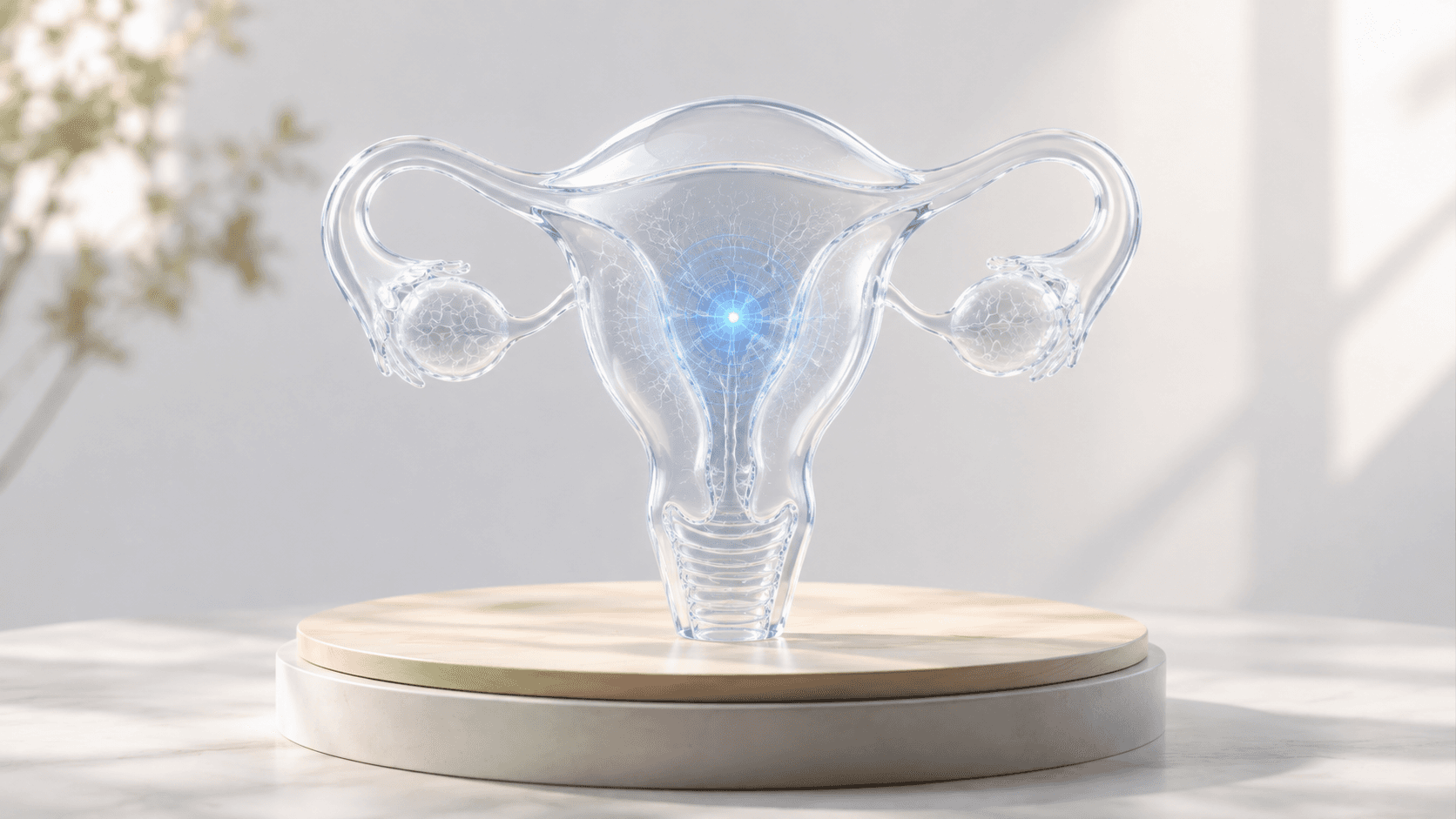
Some women’s health conditions can cause persistent pain, pressure symptoms, bleeding abnormalities, or discomfort that affects everyday life. At Nordic Focal Therapies, we provide specialist assessment for selected conditions already treated within image-guided medicine, using advanced imaging to clarify the diagnosis and identify personalised care options.
Conditions We Evaluate
Uterine Fibroids (Myomas)
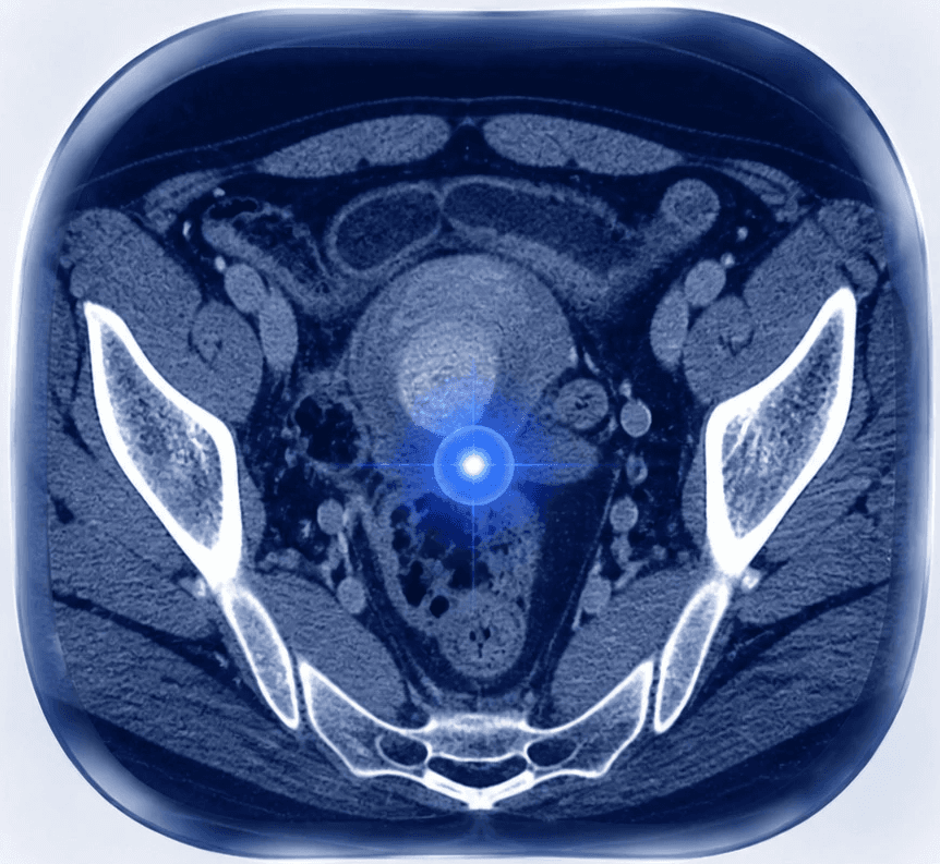
Uterine fibroids, also known as myomas, are non-cancerous growths arising from the muscular tissue of the uterus. They are common and may cause heavy or prolonged menstrual bleeding, pelvic pressure, lower abdominal pain, increased urinary frequency, or a sense of fullness. Treatment decisions depend on the size and location of the fibroids, symptoms, age, and personal goals.
Endometriosis-Related Lesions
Endometriosis-related lesions can occur when tissue similar to the lining of the uterus grows outside the uterus. In some patients, localised or scar-related abnormalities may cause persistent pain, tenderness around a previous surgical scar, or symptoms that change with the menstrual cycle. Imaging helps specialists understand the location and characteristics of the affected tissue.
Pelvic Venous Congestion
Pelvic venous congestion is a vascular condition involving abnormal veins in the pelvis. It may contribute to chronic discomfort, pressure sensations, or pain affecting daily activities. Careful imaging is important for evaluating the vascular anatomy and deciding whether an image-guided vascular treatment may be appropriate.
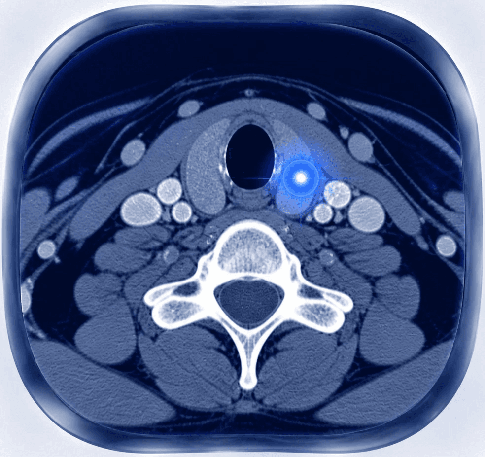
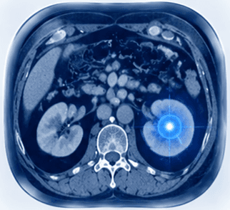
A specialist assessment combines symptoms, medical history, and imaging findings. Ultrasound is often used to evaluate pelvic conditions and vascular abnormalities in real time. MRI can provide detailed soft tissue information for endometriosis-related lesions and complex anatomical areas. CT may provide further anatomical detail and support treatment planning when needed.
Image-Guided Treatment Options
For selected symptomatic conditions, image-guided approaches may offer a minimally invasive alternative to more extensive procedures. Depending on the diagnosis and individual situation, options may include image-guided vascular treatments, microwave ablation, cryoablation, or image-guided biopsies when tissue diagnosis is needed. Every treatment decision is individualised and based on imaging, symptoms, previous treatments, and personal goals.
Uterine Artery Embolisation (UAE)
Uterine artery embolisation (UAE) is a minimally invasive image-guided treatment for selected symptomatic uterine fibroids. Using X-ray guidance, a specialist places a small catheter into the arteries supplying the fibroids and delivers tiny particles to reduce their blood supply. Over time, the fibroids shrink, which can help reduce heavy bleeding, pelvic pressure, pain, and urinary symptoms. UAE is considered after a specialist review of symptoms, imaging findings, fibroid location, and personal treatment goals.
When to Seek Specialist Assessment
A specialist review may be appropriate if you have uterine fibroids causing symptoms, persistent pain associated with an endometriosis-related lesion, suspected pelvic venous congestion, an uncertain finding on imaging, or questions about minimally invasive treatment options. A detailed review can help clarify the diagnosis and identify the most appropriate next step.
Frequently Asked Questions
Do all uterine fibroids require treatment?
No. Many fibroids can be monitored. Treatment is considered when symptoms, growth, or functional problems affect quality of life.
Can women’s health conditions be treated without surgery?
Some selected conditions may be suitable for minimally invasive image-guided procedures. Suitability depends on the diagnosis, anatomy, symptoms, and individual clinical situation.
What imaging is needed for treatment planning?
The imaging needed depends on the condition and proposed treatment. Ultrasound is commonly used to assess uterine fibroids and pelvic blood vessels in real time. MRI can provide detailed information about fibroid location, endometriosis-related lesions, and surrounding soft tissues. CT may be used when additional anatomical detail is required. For uterine artery embolisation, specialists review the relevant imaging to map the fibroids and blood supply before planning the procedure.